 The Importance of the Pelvic Sonogram
 by Jeffrey Dach MD
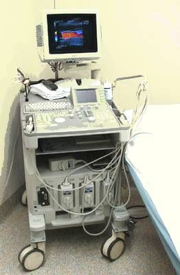
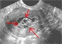
A 56 year old female patient had an episode of abnormal post-menopausal vaginal bleeding for which a pelvic sonogram was recommended. The sonogram showed a cystic swiss cheese appearance of the endometrium which looked like the image at the upper right (red arrows) Above Left Image: Ultrasound Machine Courtesy of Wikimedia
Example of Abnormal Endometrium on Pelvic Sonogram
 Left Image: Pelvic sonogram showing the abnormal endometrium (Red Arrows).
 Notice the small black areas which indicates small fluid collections.
The abnormal findings required two procedures. First, an endometrial biopsy was performed and later, a D and C (Dilatation and Curettage) for a complete removal of the abnormal endometrial tissue. Fortunately, the pathology report was favorable, showing benign endometrial hyperplasia and polyps. There was no evidence of endometrial cancer.


Above Diagram on the left shows the uterine outer muscular layer also called the myometrium. Image on tight shows the inner uterus, also called the endometrium. This is the layer that is shed every month during the bleeding period.

Uzzi Reiss MD, author of the book,, states that it is important to routinely include a pelvic sonogram for all new patients starting bio-identical hormones.
Dr. Uzzi Reiss’s reasons for a baseline pelvic sonogram:
1) A pelvic sonogram should be added to the manual pelvic exam because the old gyne pelvic exam is a 1950's standard of care. The new standard of care for the year 2000 is a pelvic sonogram.
2) The annual pelvic exam is a useful exam, but is incomplete. It provides a pap smear, visualization of the cervix, and vaginal mucosa. It cannot provide much information about the uterus and ovaries unless these organs are grossly enlarged.
3) The pelvic sonogram provides much more detailed information about the uterine size, shape and consistency, endometrial thickness, and presence or absence of fibroids, masses, or polyps in the uterus. Small masses and abnormalities can be seen on pelvic sonogram which would never be detected on manual pelvic exam.
4) If endometrial thickness is greater than 5 mm, then endometrial biopsy is usually performed to rule out endometrial cancer.
The image shows endometrial cancer. The small image at the lower lower left is a normal uterine cavity, and the larger image on the right shows a white cauliflower mass growing into the endometrial cavity. This is endometrial cancer.

5) Pelvic sonogram is more sensitive than pelvic exam for detection and evaluation of ovarian, adnexal masses, cysts, and free fluid in the pelvis.
6) Post-menopausal bleeding may occur from fluctuating hormone levels when first starting a bio-identical hormone program. Abnormal vaginal bleeding requires a pelvic sonogram to evaluate the cause of the bleeding. Having a prior baseline sonogram for comparison aids in the interpretation, and can avoid unnecessary procedures.


Above Image: Pelvic Sonogram on the left shows small black spots along the outer margin of the ovary, these are multiple small benign ovarian cysts. The image on the right shows a tubo-ovarian abscess. These findings would be difficult or impossible to detect with manual pelvic exam.
The sonogram can easily determine the cause of abnormal vaginal bleeding: Causes Include the following list:
Diffuse Endometrial hyperplasia (overgrowth)
Diffuse Endometrial cancer
Endometrial polyps
Submucosal fibroids
Focal Endometrial hyperplasia
Focal Endometrial cancer
Hormone imbalance
Simply endometrial atrophy (undergrowth)
Uterine fibroids can cause bleeding and this is what uterine fibroids look like:


Above Image: Left image is an anatomic diagram and the right image is a sagittal MRI Scan showing uterine fibroids (black spots on the MRI). On the MRI, the sacrum is at the right, and the anterior abdominal wall is at the left. The triangular shaped bladder contains white urine below and to the left of the enlarged fibroid uterus.
Sometimes bleeding can be caused by benign endometrial polyps as shown below (white arrows):

If endometrial thickening (greater than 5 mm) or other abnormality is seen on a sonogram, then endometrial biopsy is usually done. The pelvic sonogram below illustrates the endometrial stripe which is outlined with the yellow line on the right, and the blue line outlines the uterus.


The Endometrial stripe should be less than 5 mm in thickness.
Here is an example of a thickened endometrial stripe suspicious for cancer which requires endometrial biopsy for diagnosis: On the left is a trans-abdominal sonogram, and on the right is a trans-vaginal sonogram showing more detail.


The endometrial biopsy is a 10 minute gyne office procedure in which an instrument is inserted through the endocervical canal and a small sample of the endometrial lining is obtained for pathology analysis. If the pathology is abnormal, a follow up procedure called a D and C (dilatation and curettage) is done to obtain a larger sample, and to remove the entire lesion which is also sent for pathology analysis. This diagram shows the endometrial biopsy instrument in place inside the endometrial cavity performing a biopsy:

Endometrial Biopsy
For More information on Endometrial Biopsy, Click Here
There are no known harmful effects associated with the medical use of sonography which has not been shown to cause any harm or any adverse outcomes.
The transabdominal sonogram is usually the initial exam. This is done with a full bladder to provide an acoustic window for the transducer which is placed on the abdomen to obtain images of the uterus and ovaries. If needed, a more sensitive transvaginal sonogram is done with a special transvaginal transducer. This provides high resolution images of the uterine contents and ovaries.
Why You Need A Pelvic Sonogram
I hope the above discussion has convinced you of the importance of the pelvic sonogram, and why we ask that all post Menopausal women have a Baseline Pelvic Sonogram before starting our Bio-Identical Hormone Program.
A pelvic exam is 1950's standard of care. A pelvic sonogram is newer technology and current standard of care for 2000-2008.
Call Us to Schedule Your Pelvic Sonogram
Still haven't had your pelvic sonogram? To schedule your pelvic sonogram, call the office at 954-792-4663 and we will arrange it for you.
Call Us if you Need a Gyne Doctor Referral
Are you in between doctors, and looking for a good gyne doctor? We know most of the gyne doctors in the community and can refer you to someone suitable for your needs. Call the office and you can select a gyne doctor from our own list of top doctors.
Articles with Related interest
The importance of Bioidentical Hormones
Jeffrey Dach MD
7450 Griffin Rd Suite 180/190
Davie, FL 33314
Phone: 954-792-4663
Blog
References:
1) Ultrasound in the Evaluation of Abnormal Vaginal Bleeding Massachusetts General Hospital Department of Radiology
2) Ultrasound of ovarian cancer Massachusetts General Hospital Department of Radiology
3) Uterine Fibroid Embolization Massachusetts General Hospital Department of Radiology
Jeffrey Dach MD
7450 Griffin Road Suite 190
Davie, Florida 33314
954-792-4663
http://www.drdach.com/
http://www.naturalmedicine101.com/
http://www.truemedmd.com/
http://www.bioidenticalhormones101.com/
Disclaimer click here: http://www.drdach.com/wst_page20.html
The reader is advised to discuss the comments on these pages with
his/her personal physicians and to only act upon the advice of his/her
personal physician. Also note that concerning an answer which appears as
an electronically posted question, I am NOT creating a physician —
patient relationship.
Although identities will remain confidential as much as possible, as I can not control the media, I can not take responsibility for any breaches of confidentiality that may occur.
Copyright (c) 2014 Jeffrey Dach MD All Rights Reserved
This article may be reproduced on the internet without permission,
provided there is a link to this page and proper credit is given.
FAIR USE NOTICE: This site contains copyrighted material the use of which has not always been specifically authorized by the copyright owner. We are making such material available in our efforts to advance understanding of issues of significance. We believe this constitutes a ‘fair use’ of any such copyrighted material as provided for in section 107 of the US Copyright Law. In accordance with Title 17 U.S.C. Section 107, the material on this site is distributed without profit to those who have expressed a prior interest in receiving the included information for research and educational purposes.
Serving Areas of: Hollywood, Aventura, Miami, Fort Lauderdale, Pembroke Pines, Miramar, Davie, Coral Springs, Cooper City, Sunshine Ranches, Hallandale, Surfside, Miami Beach, Sunny Isles, Normandy Isles, Coral Gables, Hialeah, Golden Beach ,Kendall,sunrise, coral springs, parkland,pompano, boca raton, palm beach, weston, dania beach, tamarac, oakland park, boynton beach, delray,lake worth,wellington,plantation